
All About Accessory Navicular Bones
What will this section cover?
This section will inform you about accessory navicular bones, associated syndromes, and symptoms of disorders so you can take proactive measures towards physical well-being.
What is the accessory navicular bone?
The accessory navicular bone (ANB), also known as the os tibiale externum or os naviculare accessorium, is a small extra bone or piece of cartilage located on the inner arch of the foot. It develops within the posterior tibial tendon (which supports the arch) and is adjacent to the navicular bone. While it is a normal anatomical variant, the ANB can sometimes cause issues.
How does an accessory navicular bone form?
The accessory navicular bone (ANB) results from an anomaly during bone formation. Specifically, it occurs when an extra bone or piece of cartilage fails to detach from the navicular bone in the foot or when the navicular bone doesn't fully ossify during embryonic development. The ANB is congenital and present in approximately 10–14% of the population.
How can one identify an accessory navicular bone?
While most people with an accessory navicular bone (ANB) experience no symptoms, others may experience pain, swelling, and tenderness around the arch of the foot. These symptoms can worsen with pressure from walking or running, and some individuals may develop flat feet or arch deformities. Additionally, physical signs of inflammation or a bony lump under the skin may be noticeable, especially if the ANB is classified as type 2 or 3. The most common way to confirm the presence of an ANB is through medical imaging techniques, such as X-rays, MRIs, or CT scans.
Are all accessory navicular bones the same?
No, there are three primary types of accessory navicular bones, and not all of them cause problems.

Anatomy of the Accessory Navicular Bone and Surrounding Structures
The accessory navicular bone (ANB) is located medially (on the inner side) of the foot, where the posterior tibial tendon attaches to the navicular bone. It is embedded within the posterior tibial tendon and can vary in size and shape (as described in the classifications below). The ANB is adjacent to the navicular bone, a tarsal bone that plays a key role in forming the medial longitudinal arch and serves as a critical connection point between the forefoot and hindfoot. The posterior tibial tendon is an essential tendon in the foot. It originates from the posterior tibialis muscle in the lower leg, runs behind the medial malleolus (inner ankle bone), and inserts onto the navicular tuberosity. In individuals with an ANB, the posterior tibial tendon may attach to the ANB instead of the navicular bone, potentially leading to painful conditions.
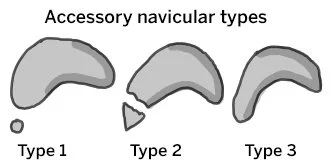
Different types of Accessory navicular bones
There are three main types of ANBs, classified based on shape, size and attachment location. To see smaller subclassifications of ANBs, check out the “Additional Resources” section.
-
A small sesamoid bone, located inside the posterior tibial tendon, is about 2-5 mm in size. It is not connected to the navicular bone, and since it is embedded within the posterior tibial tendon, it is usually asymptomatic.
-
A larger, triangular or heart-shaped accessory bone, about 7-12 mm in size, is connected to the navicular bone by a fibrocartilaginous or hyaline cartilage synchondrosis. It is more prone to symptoms due to irritation, inflammation, or mechanical stress. Type II is often associated with flat feet and may require conservative or surgical treatment if symptomatic.
-
An accessory bone that is completely fused with the navicular, forming an enlarged navicular bone. It may cause a bony protrusion on the medial side of the foot, leading to pain and discomfort from shoes. While it is less likely to cause pain than Type II, it can still cause discomfort in some individuals.
Accessory Navicular Bones Are Linked to a Number of Foot Conditions, Common Disorders Are Included Below:

Accessory Navicular Bone Syndrome (ANBS)
A painful syndrome that occurs due to injury, excessive activity, or footwear irritation. Common symptoms include pain, inflammation, swelling, or redness on the inner side of the foot. Individuals with ANBS may also experience a prominent bony lump next to the navicular, difficulty walking, or a flat foot deformity. Symptoms can worsen with physical activity or weight-bearing. ANBS is most common in individuals with a Type II ANB and typically appears during the teenage years.
Posterior Tibial Tendon Dysfunction (PTTD)
Normally, the posterior tibial tendon (PTT) inserts into the navicular bone; however, it can occasionally insert abnormally into the accessory navicular bone. This leads to a weaker attachment, making the PTT more prone to strain and dysfunction. Common symptoms include inflammation of the PTT and progressive flatfoot deformity. The disorder is also associated with ANB pain, and when combined with posterior tibial tendon dysfunction (PTTD), the foot may become more painful, swollen, and immobile.


Flatfoot (Pes Plantus)
The presence of an accessory navicular bone (ANB) is associated with the development of flat feet, particularly flexible flatfoot. The posterior tibial tendon is essential for stabilizing the medial longitudinal arch of the foot. With the presence of an ANB, particularly Type II, the biomechanics of the tendon can be affected, interfering with normal arch formation and function. A weakened or irritated tendon leads to progressive arch flattening, pain, inflammation, posterior tibial tendon dysfunction (PTTD), and mechanical instability. Those with a Type II ANB or pre-existing flatfoot deformities are most at risk.
Medial Tibial Stress Syndrome (Shin Splints)
Since the accessory navicular bone (ANB) is embedded in or attached to the posterior tibial tendon, which helps maintain the foot's arch, it can contribute to excessive foot pronation (inward rolling). Overpronation places excessive stress on the shin muscles (tibialis posterior, tibialis anterior), leading to muscle imbalances, fatigue, and improper shock absorption. This causes strain and inflammation along the tibia. The added stress increases the risk of tendon overuse, fatigue, and microtears, all of which can contribute to shin splints.


Stress Fractures or Navicular Bone Irritation
When the accessory navicular is exposed to excessive stress, it can cause micro fractures, tiny cracks in the bone, or chronic irritation. If the posterior tibial tendon is weak or inflamed, other parts of the foot are forced to compensate, shifting excess load to the ANB and increasing the risk of stress fractures over time. Repetitive high impact activities pose, the ANB absorbs stress, leading to progressive micro damage. The ANB is a bony prominence, meaning its subject to direct impact and pressure from footwear or external forces. Continues friction or compression from tight shoes, or foot strikes during running can contribute to bone irritation and stress fractures.
How to Reduce the Likelihood of Complications from an Accessory Navicular Bone?
-
Supportive footwear with built-in arch supports helps maintain proper foot alignment and reduces stress on the accessory navicular bone (ANB). A cushioned shoe can minimize stress traveling up the foot and decrease the risk of shin splints. Additionally, stability-focused shoes limit inward movement of the foot, easing pain from the ANB. Shoes with a wide toe box may also reduce strain on the bone and improve gait.
-
Many people with ANBs have flat feet or pronation. Orthotics provide arch support, preventing excessive pronation and reducing strain on the tendon and surrounding tissues. Cushioned orthotics absorb shock, reducing the force transmitted to the ANB and surrounding structures. Orthotics offer structural support, reducing muscle fatigue and the risk of conditions like shin splints, posterior tibial tendinitis, and plantar fasciitis. Over time, orthotics improve foot posture and function, helping to reduce future foot problems.
-
Strengthening the intrinsic foot muscles (such as the flexor digitorum brevis and abductor hallucis) through toe isolation, barefoot walking, short foot exercises, and other movements helps maintain proper arch shape. Improving posterior tibial tendon strength through arch lifts can reduce tendon overload and discomfort. Supporting foot and ankle stability with balancing exercises and ankle band resistance can strengthen the peroneal and tibialis anterior muscles, preventing overuse injuries. Additional activities like calf raises and toe taps can reduce impact forces on the foot and shin. Other targeted exercises are listed in the "Additional Information" section.
-
Stretching the lower leg and foot muscles helps reduce tension, improve flexibility, and prevent excessive strain on the posterior tibial tendon and ANB. Use the posterior tibial tendons stretch or seated foot stretch to support proper tension function. Improving foot and ankle flexibility through calf and soleus stretches promotes foot mechanics and reduces abnormal stress. Tight calf muslces can contribute to overpronation and ANB stress. The Plantar Fascia stretch and Tibialis Anterior stretch, help restore proper foot alignment. Calf foam rolling and toe raises can reduce shin splint risks. To encourage better shock absorption and reduce impact force, ankle circles and dynamic Achilles stretches are solid options. More stretches are included in the “Additional Resources” segment.
-
Modifying activities can reduce stress on the accessory navicular bone (ANB), prevent pain and inflammation, and minimize the risk of long-term complications. Limit high-impact activities like running, jumping, and prolonged walking if pain occurs, and incorporate rest days between intense activities. To prevent overloading the posterior tibial tendon, it is recommended to use interval training and cross-train with low-impact activities. Modify exercise techniques to avoid direct pressure on the ANB and always listen to your body. Seek professional guidance if painful symptoms persist.